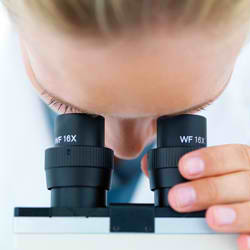
How little things can you see in microscopes? The light itself sets a limit. But there are ways to trick the light on. A Polish student has made the economy version of a super microscope.
The laser microscope as master student Joanna Oracz at the University of Warsaw have made, similar to the commercial variety, but also has several properties that are valuable to researchers.
- The difference is that we can control all the parameters, not only the strength of the laser beams, but also the size of the rays, the properties of the pulses, polarization, time management and so on.
- Besides, this system is much cheaper than the commercial, she emphasizes.
See without killing
The microscope will be primarily used to study living cells. They are quite small, a hundredth of a millimeter from beginning to end. But the big that an ordinary microscope can see them.
More difficult is it when scientists will zoom into the cell nucleus. A solution to see the details, is to use an electron microscope.
But when they enter the cell with formaldehyde to stiffen it up, freeze it down with liquid nitrogen and cover it with lead or uranium for better contrast.
When scientists can get a very sharp image of a very, very dead cell nucleus. But if you want to study life, there is always an advantage that it is alive.
Like rings on a pond
Conventional microscopes that use visible light does not kill cells. Is it not just to build a very good optical microscope?
I'm afraid not. For the problem to the light microscope is not in the lenses. It is in the light.
Light is waves. And when the light waves breaking against the lens of the microscope, is something of the same as when the rings spreading on a pond.
If the blubber
As long as the waves are waves, above the surface, the rings are smooth and pretty. But when they hit the rocks on the shore, broken rings up. Waves collide with the waves, and stock patterns.
The same thing happens when light waves hit the lens of the microscope. Diffraction transforms a bright spot to a lysblubb with rings around.
No matter how good the lens is, set a lower limit to how sharp the image will be.
Sharpen the light!
In 1957, took the American scientist Marvin Minsky patented a new way to use the light on. In his microscope was not only the lenses used to focus the object that should be enlarged. It was used to focus the light source, the light shining on the object.
A more modern version has replaced a conventional lamp with a laser. Laser light has a very specific color, so that all light waves fluctuate in time.
When it is focused through a lens, hits the object in a tiny flare.
Vibrant and luminous
But the point of this laser light is not illuminating the object. It is to get the object to illuminate itself.
If the object is a living cell, it is inserted with a fluoriscerende fabric. This substance will light when the laser hit it, but with a different color. With a color filter may let through fluorescence, and laser light.
And the best part is that the cell survives.
As 3D TV
But how can you create an image with a small bright spot? By letting the bright spot sweeping over the object. Then build the picture up like an old fashioned TV screen, line by line.
Another advantage of this microscope is that the laser light is focused at a certain distance from the lens.
This means that parts of the cell that is higher or lower than this distance, not made by a bright spot, but a field of light that is smeared out over a larger area. The brightness and the fluorescence is much weaker.
On the way, the only part of the cell at a particular height scanned. By running multiple scans with different focuses, different depth levels depicted.
Together they provide a three-dimensional, very sharp image of the living cell.
Could be better
Marvin Minsky microscope called a konfokalt scanning microscope. But even this microscope has its limitations.
Bright spot that scans of the living cell is certainly much less than diffraksjonsblubben in a conventional microscope.
But because the light source is focused by a lens, too, this lens produce tiny diffraksjonsmonstre and lubricating bright spot beyond.

In 1999, managed the Romanian physicist Stefan Hell to get it konfokale scanning microscope to sharpen up even more. To achieve this, he used two different lasers.
Very simply explained as pursed a ring of quenching laser light up around the foksuerte laser spot that gets fluorescence. Thus, the bright spot sandwiched together, and are up to twelve times less.
The method is called in English Stimulated Emission Depletion microscopy copy (PLACE). Stimulated emission is, laser, and depletion refers to quench the function of one laser.
For the first time, scientists can see the individual organellene and large molecules in a living cell. Details down to a little over five nanometers is depicted in the PLACE-microscope as Stefan Hell invented.
Even sharper
- In the future we would like to study the physical properties of our system, and find the optimal parameters to achieve the best resolution.
Microscopes can apparently shape up even more.


